



Could an MRI Detect Alzheimer’s Years Before Memory Loss? New Stanford Research Offers Hope
November 5, 2026 | VietEcho Health
Imagine visiting your doctor while your memory is still perfectly normal, yet a specialized brain scan reveals subtle changes associated with a higher risk of developing Alzheimer’s disease years later.
That possibility may be moving closer to reality.
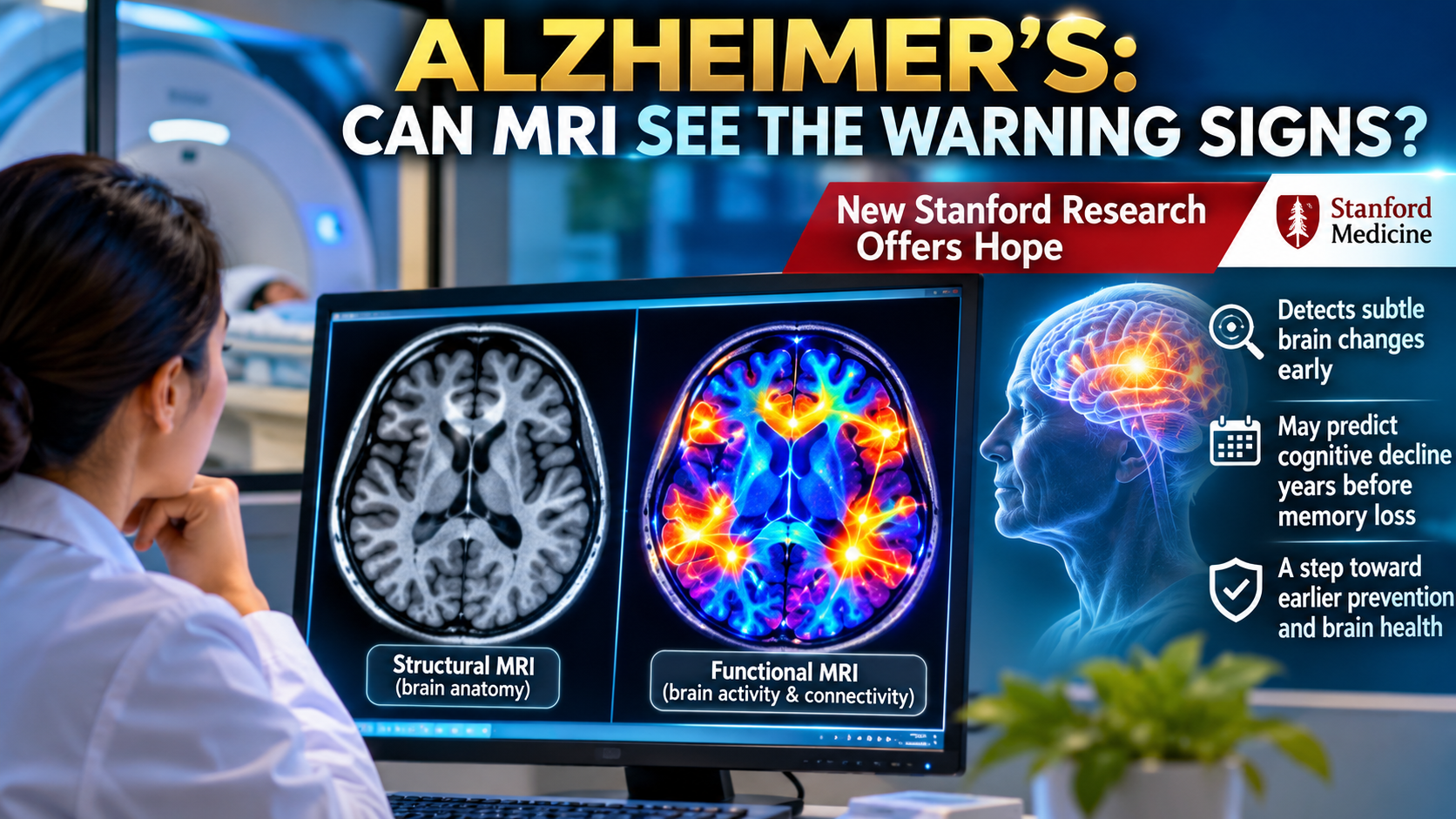
A new study led by researchers at Stanford Medicine, published November 4, 2026, in Science Advances, suggests that patterns of communication between different brain regions may help identify people vulnerable to future cognitive decline—even before they experience noticeable memory problems.
The discovery could eventually open new opportunities for earlier monitoring and preventive research.
1. What Did Stanford Researchers Discover?
The researchers examined how different regions of the brain communicate with one another while a person is resting.
They identified two contrasting patterns of brain activity.
One pattern resembled the activity found in people diagnosed with Alzheimer’s disease. Another resembled the activity of unusually healthy older adults who maintain exceptional memory performance.
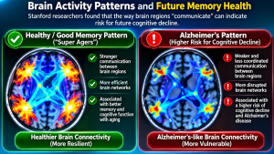
Interestingly, these patterns could also be detected in younger adults who had no apparent cognitive impairment.
People whose brain activity more closely resembled the Alzheimer’s pattern were more likely to experience cognitive decline during follow-up.
Those whose brain activity resembled the exceptionally healthy aging pattern tended to maintain better cognitive performance.
The findings suggest that changes in brain communication may provide clues about future cognitive health.
2. What Are “Super Agers”?
Some people remain mentally sharp well into their late 70s, 80s, and beyond.
Researchers often refer to individuals with unusually preserved memory as super agers.
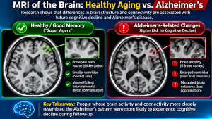
In this study, super agers were adults aged 75 or older whose memory performance was comparable to that of people around age 45.
Scientists compared brain scans from 180 super agers with scans from 110 individuals diagnosed with Alzheimer’s disease.
They discovered distinct patterns of communication between brain regions.
Using these differences, researchers developed a scoring system capable of distinguishing the two groups with approximately 85% accuracy.
Importantly, this figure describes how well the model separated the two established groups. It does not mean an MRI can predict Alzheimer’s disease in healthy individuals with 85% accuracy.
3. How Does Functional MRI Detect Brain Activity?
Unlike a conventional MRI, which primarily produces detailed anatomical images, functional magnetic resonance imaging (fMRI) examines changes associated with brain activity.
Functional MRI uses a technique called the blood-oxygen-level-dependent, or BOLD, signal.
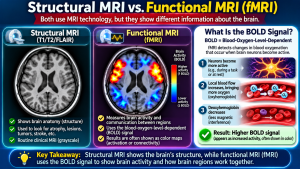
When brain cells become active, local blood flow and oxygenation change. These changes produce measurable variations in the MRI signal.
In this study, participants underwent resting-state functional MRI.
They did not need to perform complicated memory tests inside the scanner.
Instead, researchers examined how activity fluctuated across different brain regions while participants rested.
By analyzing these patterns, scientists could estimate how strongly different parts of the brain were functionally connected.
These communication patterns may reveal changes that are not visible on ordinary structural MRI images.
4. Could These Changes Appear Years Before Symptoms?
This was one of the study’s most important findings.
After developing their model, the researchers evaluated more than 1,300 adults between ages 55 and 70 who showed no signs of cognitive impairment when their brain scans were obtained.
At the beginning, participants with different brain activity patterns performed similarly on cognitive tests.
However, differences emerged during follow-up.
Individuals with more Alzheimer’s-like brain activity experienced faster declines in memory, attention, and executive function.
They were also more likely to develop Alzheimer’s-related changes.
Some participants had follow-up information extending as long as 12 years, although most were followed for approximately five to six years.
The findings suggest that resting-state brain activity may contain early warning signals of cognitive decline.
5. What Makes This Discovery Different?
Much Alzheimer’s research focuses on two proteins:
Amyloid-beta: A protein that can accumulate in plaques between brain cells.
Tau: A protein that can form abnormal tangles inside nerve cells.
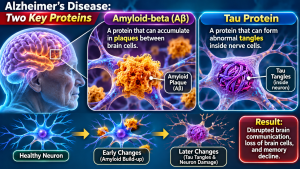
These changes are strongly associated with Alzheimer’s disease.
However, researchers are interested in finding warning signs that may emerge even earlier.
The Stanford team developed a measurement called the Alzheimer’s Resilience-Vulnerability Connectome (ARC) score.
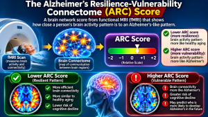
This score reflects whether an individual’s brain communication pattern more closely resembles that of super agers or people with Alzheimer’s disease.
In the research datasets, the score performed comparably to or better than several established risk indicators for predicting later cognitive decline.
Combining the ARC score with existing biomarkers improved prediction further.
6. Does This Mean Everyone Should Get a Brain MRI?
Not yet.
Although the results are encouraging, the technology remains experimental.
The ARC score is not an established clinical diagnostic test for Alzheimer’s disease.
A routine brain MRI cannot reliably determine whether someone will develop Alzheimer’s years later.
Researchers must validate the findings in additional populations and determine whether the scoring system can improve clinical decisions.
It is also important to remember that having an Alzheimer’s-like brain activity pattern does not guarantee that a person will develop dementia.
Risk is not destiny.
7. Could Scientists Eventually Protect the Brain?
One particularly exciting possibility is that brain activity patterns might be modifiable.
Researchers are interested in whether interventions such as cognitive training or transcranial magnetic stimulation could influence brain networks associated with resilience.
However, these approaches have not yet been proven to prevent Alzheimer’s disease by changing ARC scores.
The Stanford team plans further research to investigate whether cognitive training can alter these brain activity patterns.
For now, the discovery offers a promising direction for future prevention studies rather than an established treatment.
8. What Can People Do Today to Support Brain Health?
While researchers continue investigating new imaging biomarkers, several everyday habits may help support long-term cognitive health.
Regular physical activity, sufficient sleep, management of blood pressure and diabetes, social engagement, and mentally stimulating activities are important components of healthy aging.
A balanced diet rich in vegetables, fruits, whole grains, legumes, and other nutrient-dense foods may also support cardiovascular and overall brain health.

People experiencing persistent or worsening memory problems should discuss their symptoms with a healthcare professional rather than relying on experimental imaging tests.
Conclusion: A New Window Into the Aging Brain
The Stanford discovery suggests that the brain may reveal important clues about future cognitive health long before obvious memory problems appear.
By examining how brain regions communicate, researchers may eventually develop better ways to identify people at higher risk and investigate interventions before significant cognitive decline occurs.
Although more research is necessary, the findings represent an encouraging advance toward understanding why some brains remain resilient while others become vulnerable to Alzheimer’s disease.
The ultimate goal is not simply to predict memory loss, but to discover how it might someday be delayed or prevented.
Scientific source: Zhao K. et al. A Functional Connectome Identifies Vulnerability to Cognitive Decline and Alzheimer’s Disease Progression. Science Advances, November 4, 2026. DOI: 10.1126/sciadv.aed9351.
Medical disclaimer: This article is for educational purposes and does not replace professional medical advice, diagnosis, or treatment.